
#PathPointer: An Approach to Glomerular Disease in Kidney Biopsies
Summer marks the graduation of nephrology fellows and welcoming of new ones. In this post, we will provide an image-rich, pattern-based approach to glomerular disease in kidney biopsies. Many others are available elsewhere; Fundamentals of Renal Pathology, by Fogo et al is also an excellent resource.
Light microscopy
- Are glomeruli normal or abnormal? (Figure 1)
- Is there too much matrix, too many cells, or both?
- Where is the abnormality?
- Mesangium (Figure 2, 4)
- Capillary loops (Figure 3, 4, 5)
- Bowman’s space (Figure 5, 6)
- Does the abnormality involve all of the glomerular tuft (global) or part of it (segmental)? (Figure 6)
- Do the abnormalities involve all glomeruli (diffuse, >50%), or some of them (focal, <50%)?
Immunofluorescence microscopy
- Deposit type (Figure 7)
- IgG, IgM, IgA, kappa light chain, lambda light chain, complement C3, complement C1q
- Deposit location (Figure 7)
- Mesangium, capillary loop, outside glomeruli
Electron microscopy
- Deposit location (mesangium, subendothelial, subepithelial) (Figure 7)
- Deposit substructure, if present
- Glomerular basement membrane abnormalities, if present
- Supportive or unexpected findings
Integrate
- Correlate with clinical, lab, systemic features
- Degree of activity and chronicity
- Do the findings provide an anatomic etiology for the observed clinical abnormality and reason for biopsy? (If not, why not? What are the pertinent negatives?)
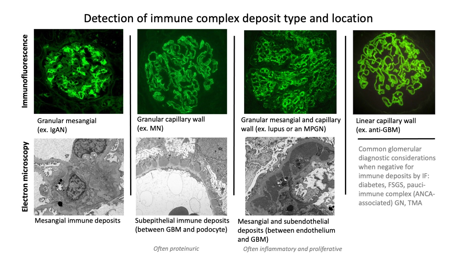
Figure 7
– Post prepared by Nicole Andeen, AJKDBlog Contributor
Leave a Reply